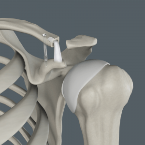
What is the Acromioclavicular (AC) Joint?
The acromioclavicular (AC) joint is one of the joints present within your shoulder. It is formed between a bony projection at the top of the shoulder blade (acromion) and the outer end of the clavicle (collarbone). The joint is enclosed by a capsule and supported by ligaments.
The AC joint is stabilized by the following structures:
- Capsular ligaments: These ligaments are called the acromioclavicular ligaments. They have superior and inferior components and resist separation of the joint in the horizontal direction.
- Extracapsular stabilizers: These are ligaments extending from a bony process of the scapula called the coracoid process to the clavicle (coracoclavicular ligaments) and the acromion (coracoacromial ligaments). These ligaments resist vertical forces from separating the joint.
- Muscular attachments: The deltoid muscle on the outside of the shoulder and the trapezius muscle in the upper back and neck also help stabilize the acromioclavicular joint.
An injury to the AC joint, particularly the ligaments, can result in instability or separation of the AC joint (shoulder separation) causing pain and discomfort and limiting shoulder function.
What is Acromioclavicular Joint Reconstruction?
Shoulder separation can usually be managed by non-surgical treatments. In cases of a severe separation of the AC joint, your surgeon may perform a surgical repair or use a tissue graft to reconstruct the damaged ligaments. An autograft (from your own body) or allograft (from a donor) of the anterior tibia or hamstring may be used. Your surgeon might also suggest artificial non-absorbable material to graft the damaged ligaments.
The goals of surgery include:
- Relief from pain
- Improved shoulder movement
- Improved strength and function
- Faster recovery in cases of athletes
Anatomic reconstruction of the AC joint helps ensures static and safe fixation with stable joint function.
Procedure for Acromioclavicular (AC) Joint Reconstruction
Acromioclavicular joint reconstruction involves the following steps:
- The procedure is performed under general anesthesia and regional anesthesia.
- A diagnostic arthroscopy may be performed to visualize the position of the tear and extent of the damage.
- An incision is made to expose the AC joint and any damaged tissue is trimmed.
- Tissue is dissected to expose the coracoid process.
- The graft is passed from under the coracoid process to a hole drilled into the clavicle and the displaced clavicle is reduced.
- Sutures are used to close the wound.
- Alternatively, a ligament extending from the coracoid process to the acromion (coracoacromial ligament) may be transferred to the end of the clavicle to reduce it in position.
- The graft ends are secured to the bone.
Following surgery, your arm is placed in a sling and your shoulder will be immobilized for a period of 6 weeks. You will then require rehabilitation to regain strength and movement of the shoulder joint and can return to your regular activities in about 6 months.
Related Topics
- Shoulder Joint Replacement
- Shoulder Arthroscopy
- Rotator Cuff Repair
- SLAP Repair
- Revision Shoulder Replacement
- Minimally Invasive Shoulder Joint Replacement
- Reverse Shoulder Replacement
- Shoulder Labrum Reconstruction
- Pectoralis Major Tears/Repairs
- Acromioclavicular (AC) Joint Reconstruction
- Shoulder Stabilization
- Proximal Biceps Tenodesis
- AC Joint Stabilisation
- ORIF of the Scapula Fractures
- Arthroscopic Frozen Shoulder Release
- Capsular Release
- Bony Instability Reconstruction of the Shoulder
- ORIF of the Clavicle Fractures
- Arthroscopic Superior Capsular Reconstruction (SCR)
- Subacromial Decompression
- Shoulder Resurfacing
- Periprosthetic Shoulder Fracture Fixation
- Ultrasound-Guided Shoulder Injections
- Intraarticular Shoulder Injection


