Knee Anatomy
The knee is a complex joint that consists of bone, cartilage, ligaments, and tendons that help in your joint’s movements.
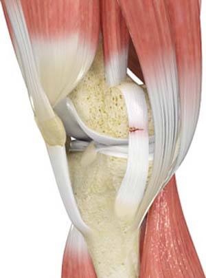
The knee is a hinge joint made up of two bones, the thighbone (femur) and shinbone (tibia). Ligaments are tough bands of tissue that connect one bone to another bone. The ligaments of the knee stabilize the knee joint. There are two important groups of ligaments that hold the bones of the knee joint together, collateral and cruciate ligaments - medial collateral ligament (MCL) and lateral collateral ligament (LCL), and anterior cruciate ligament (ACL) and posterior cruciate ligament (PCL).
Knee Injuries
Knee problems may arise if any of these structures get injured by overuse or suddenly during sports activities. Pain, swelling, and stiffness are the common symptoms of any damage or injury to the knee.
The common causes of knee injury include:
- Fracture of the femur (thighbone) or tibia and fibula (leg bones)
- Torn ligament: either anterior or posterior cruciate ligament (ACL or PCL)
- Rupture of blood vessels following a trauma that leads to the accumulation of extra fluid or blood in the joint
- Dislocation of the kneecap (patella)
- Torn quadriceps or hamstring muscles
- Patellar tendon tear
Types of Ligament Injuries
The most common types of ligament injuries include:
ACL tear
An ACL injury is a sports-related injury that occurs when the knee is forcefully twisted or hyperextended. An ACL tear usually occurs with an abrupt directional change with the foot fixed on the ground or when the deceleration force crosses the knee. Changing direction rapidly, stopping suddenly, slowing down while running, landing from a jump incorrectly, and direct contact or collision, such as a football tackle, can also cause injury to the ACL.
MCL tear
The MCL is the ligament that is located on the inner part of the knee joint. It runs from the femur (thighbone) to the top of the tibia (shinbone) and helps in stabilizing the knee. MCL injuries can result in a stretch, partial tear or complete tear of the ligament. Injuries to the MCL commonly occur because of pressure or stress on the outside section of the knee.
PCL tear
PCL injuries are very rare and are more difficult to detect when compared to the other knee ligament injuries. Cartilage injuries, bone bruises, and ligament injuries often occur in combination with PCL injuries.
Injuries to the PCL can be graded as I, II or III depending on the severity of the injury. In grade I, the ligament is mildly damaged and slightly stretched, but the knee joint is stable. In grade II, there is a partial tear of the ligament. In grade III, there is a complete tear of the ligament and the ligament is divided into two halves, making the joint unstable.
The PCL is usually injured by a direct impact, such as in an automobile accident when the bent knee forcefully strikes the dashboard. In sports, it can occur when you fall to the ground with a bent knee. Twisting injury or overextending the knee can cause the PCL to tear.
Treatments for Ligament Injuries
Immediately following a knee injury, before being evaluated by a doctor, you can initiate the R.I.C.E. method of treatment:
- Rest: Rest the knee, as more damage could result from pressure on the injury.
- Ice: Ice packs can be applied to the injured area to reduce swelling and pain. Never place ice directly over the skin. Ice should be wrapped in a towel and applied to the affected area for 15-20 minutes, four times a day for several days.
- Compression: Wrapping the knee with an elastic bandage or compression stocking can help minimize the swelling and support your knee.
- Elevation: Elevating the knee above the heart level will also help reduce swelling and pain.
It is important to seek your doctor’s advice if you hear a popping noise or feel as if your knee has given way at the time of injury and if you are unable to move your knee because of severe pain.
Related Topics
- Patellar Instability
- Multiligament Instability
- Patellofemoral Instability
- Posterolateral Instability
- Knee Arthritis
- Knee Osteoarthritis
- Knee Injury
- Knee Pain
- Anterior Knee Pain
- Meniscal Tears
- Runners Knee
- Jumpers Knee
- Unstable Knee
- Knee Sprain
- MCL Sprains
- ACL Tears
- MCL Tears
- Meniscal Injuries
- PCL Injuries
- Ligament Injuries
- Knee Fracture
- Fractures of the Tibia
- Patella Fracture
- Tibial Shaft Fracture
- Kneecap Bursitis
- Chondral or Articular Cartilage Defects
- Quadriceps Tendon Rupture
- Patellar Tendon Rupture
- Lateral Meniscus Syndrome
- Osteonecrosis of the Knee
- Knee Angular Deformities
- Osteochondral Defect of the Knee
- Articular Cartilage Injury
- Goosefoot Bursitis of the Knee
- Iliotibial Band Syndrome
- Bowed Legs
- Recurrent Patella Dislocation
- Osteochondritis Dissecans of the Knee
- Chondromalacia Patella
- Patellar Tendinitis
- Knee Sports Injuries
- Multiligament Knee Injuries
- Women and ACL Injuries
- Medial Meniscus Syndrome
- Tibial Plateau Fracture
- Tibial Eminence Fractures
- Loose Bodies in the Knee
- Osgood Schlatter Disease
- Patellar Dislocation/Patellofemoral Dislocation